$29,900.00
Fiber photometry is a technology to detecting the activity of neurons in the brain nucleus of freely moving animals. It sums up the overall fluorescence of neurons expressing a genetically encoded calcium indicator(GECI) or neurotransmitter probes.
The fiber photometry system records changes in the fluorescence intensity of neurons in a specific brain area to reflect neuronal population activity. In the study of neural circuits, the fiber photometry system can perform long-term stable monitoring of the neurons of freely moving animals, and explore the correlation between neural activity and animal behavior.

RWD is a global leader in the design and manufacturing of advanced research and laboratory equipment. Specializing in high-precision instruments for neuroscience, behavioral science, and pharmacology.


R821 TriColor Multichannel Fiber Photometry System has three excitation light sources, 410nm, 470nm and 560nm, of which 410 is used to acquire reference signal and eliminate noise. The system can record signal of green fluorescence indicator like GCaMP and dLight or neurotransmitter probe and red fluorescence indicator like RCaMP, jrGECO1a or neurotransmitter probe.
Fiber photometry calcium imaging is becoming increasingly popular in neuroscience research and, based on its advantages, is now an indispensable tool for the real-time detection of neural signals.
Product Parameter | Details |
|---|---|
Wavelength of excitation light | 410nm 470nm 560nm |
Power | Min 0µW, Max≥100µW, adjustable with an accuracy of 0.1µW |
Number of channels | 9 |
Frame rate of fluorescent sampling | Max 250fps |
Digital signal interface | 4 Input /4Output |
Multiple signal input & output ports | USB 3.0 (software control), 4 digital Signal input, 4 Digital signal output, CHAS (Grounding Interface), Optogenetics(Model R821) |
Signal output | Output frequency 0-500Hz, adjustable output pulse width and duration |
Marking | Manual marking (10), automatic marking (4), ROI marking (9) |
Behavior camera | 1920*1080(30fps) 1280*720(60fps) Switchable among multiple frame rates of resolution |
Compatible | Compatible with optogenetics for recording and stimulation at the same site |
Item | Qty | Specifications | Description |
|---|---|---|---|
Fiber Photometry Main Device | 1 | Includes: Host, power cord, 3 USB cables, USB expansion interface, software U disk | Dimension: 31.5 cm x 30.3cm x 11.1cm |
Software | 1 | Includes pre-installed software, I5-10500H/16G/ 500G/WIN10(1920*1080) | Integrated software: Data acquisition and analysis |
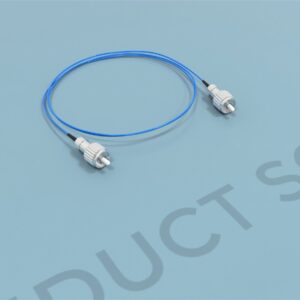
Optical fiber | 1 | Low Autofluorescence Fiber-optic Patch Cords 200um/ 0.37NA/2m,Ф1.25mm or Ф2.5mm | Small and flexible: minimal tissue damage |
Fiber Cannula sleeves | 1 | Black Ceramic Sleeves, Φ1.25mm or Φ2.5mm | -- |
Behavior Camera | 1 | Record video of animal behavior and identify animal tracks,USB3.0,3M | Flexible viewing of behavior videos. Simultaneously data in vivo animals. |
Behavior Camera bracket | 1 | Adjustable height range0.8-1.5m,Rotation Angle 360° | -- |
Photobleaching device | 1 | FC/PC Patch Cord photobleaching machine(R810-1) | Reduce autofluorescence interference |
U disk | 1 | Software key(Not for analysis function) | Back up drivers and software |
The implanted fiber optic probe is small and flexible and resulting in minimal tissue damage and allows recording from multiple brain regions simultaneously.
Bonsai software and MATLAB programming are not required. Data analysis includes data clipping, bleaching correction, smoothing, movement correction, event heat map, peak statistics, and area under curve and heat map of behavior trajectory.
4-input and 4-output interface for easy connection with other equipment such as optogenetics and electrophysiology for the closed-loop study of stimulation and recording.
The software can synchronize and mark multiple special behavioral events or external input signals during the experiment.
Support up to 9 channels, suitable for simultaneous experiment of multiple animals or multiple brain locations
Dual highly sensitive detectors enabling independent and sequential detection to avoid interference of fluorescence excitation and detection, acquiring more accurate signal. The 410nm light source can be used to reflect the background noise signal, thus ensuring the acquisition of true fluorescence data.
Fiber photometry emerges as a sophisticated technology meticulously designed for the precise detection of neural activity within the brain nuclei of unrestrained animals. This method entails the amalgamation of fluorescence emitted by neurons expressing genetically encoded calcium indicators (GECI) or neurotransmitter probes. (Adelsberger, 2005).
The evolution of fiber photometry calcium imaging spans nearly a decade, earning commendation from diverse laboratories for its rigorous scientific merit. Its application extends to the systematic investigation of regulatory mechanisms governing animal behavior. One reason for the growth of this technique is the ongoing development of biosensors that measure general cellular activity, or the activity of specific neurotransmitters such as glutamate, GABA, acetylcholine, serotonin, dopamine, norepinephrine, and orexin (Bruno et al., 2021).
Fiber photometry constitutes an optical methodology wherein light serves as a stimulus to initiate and quantify variations in fluorescence arising from structural alterations in an expressed biosensor. Briefly, light of a specific wavelength for excitation is transmitted through an implanted optical fiber, and the ensuing fluorescence is conveyed back via the same fiber to a photodetector. Subsequently, a digital optical intensity signal is generated, hypothesized to depict the proportional quantity of the target-bound sensor situated at the distal end of the fiber (Li et al., 2019).
The detected signal emanates from the tissue surrounding the fiber tip, with a spatial extent ranging from 50 to 400 um, thus constituting a regional or ‘bulk’ measurement. Owing to the genetic encoding of biosensors, their expression can be directed toward specific circuits and/or cell types, where stability may endure for extended durations, spanning weeks to months.
This protracted temporal capacity distinguishes fiber photometry from other in vivo techniques, enabling repetitive recordings over considerable time frames (Gunaydin, et al., 2014). Notably, this methodology has facilitated unprecedented insights into the correlation between population activity within specific cell groups and various facets of intricate behaviors, including but not limited to movement, memory, motivation, appetitive and aversive learning, among others.
As remarked by Simpson et al., 2023, Fiber photometry has gained popularity for in vivo monitoring of neural signals in behaving animals due to its practical advantages. In comparison to other methods like electrophysiology, it offers signals with molecular and cellular specificities, higher spatial resolution, and much higher temporal resolution. It outperforms microdialysis in terms of temporal resolution and can provide concurrent recordings of dopamine, revealing differences in observable temporal dynamics. Additionally, photometry is more sensitive for certain analytes/environments compared to cyclic voltammetry and allows access to molecules without electrochemical methods. Its practical benefits include less invasive surgical procedures, flexibility with lightweight optical fibers, and the availability of cost-effective “plug-and-play” systems, making it accessible for diverse researchers to study brain-behavior relationships at scale. Furthermore, fiber photometry generates relatively low-sized and less complex raw data compared to other in vivo techniques like electrophysiology and imaging.
The integration of fluorescent reporters with fiber photometry enables the monitoring of cell-type-specific neuronal activity and the assessment of specific gene expression levels (Chen et al., 2013). This experimental approach, crucial for investigating brain functions, involves studying correlations between neuronal activity and natural animal behavior. Traditional electrophysiological recording, renowned for its high temporal resolution, has provided profound insights into neural circuit functions. However, employing light-sensitive proteins for optical tagging in electrophysiological recordings is technically challenging, sensitive to noise interference during natural behaviors, and inefficient.
Fiber photometry, leveraging genetically-encoded Ca2+ indicators like GCaMP proteins, facilitates the monitoring of genetically-defined neuron populations. Numerous research groups have utilized fiber photometry to make exciting observations of neuronal activation patterns during various behaviors, including affective reward- and punishment-related behaviors (Wang et al., 2017), feeding behaviors, social behaviors (Chen et al., 2015), arousal, and long-term learning. Moreover, fiber photometry allows for the measurement of gene expression levels, as demonstrated in a study of circadian rhythms by monitoring circadian clock gene expression using bioluminescent reporters (Mei et al., 2018).
Adelsberger, H., Garaschuk, O., & Konnerth, A. (2005). Cortical calcium waves in resting newborn mice. Nature neuroscience, 8(8), 988-990.
Bruno, C. A., O’Brien, C., Bryant, S., Mejaes, J. I., Estrin, D. J., Pizzano, C., & Barker, D. J. (2021). pMAT: An open-source software suite for the analysis of fiber photometry data. Pharmacology, biochemistry, and behavior, 201, 173093. https://doi.org/10.1016/j.pbb.2020.173093
Chen, T.-W., Wardill, T. J., Sun, Y., Pulver, S. R., Renninger, S. L., Baohan, A., Schreiter, E. R., Kerr, R. A., Orger, M. B., Jayaraman, V., Looger, L. L., Svoboda, K., & Kim, D. S. (2013). Ultrasensitive fluorescent proteins for imaging neuronal activity. Nature, 499(7458), 295+. https://link.gale.com/apps/doc/A337370689/HRCA?u=anon~1e8d50e8&sid=googleScholar&xid=109ca2eb
Chen, Y., Lin, Y. C., Kuo, T. W., & Knight, Z. A. (2015). Sensory detection of food rapidly modulates arcuate feeding circuits. Cell, 160(5), 829–841. https://doi.org/10.1016/j.cell.2015.01.033
Gunaydin, L. A., Grosenick, L., Finkelstein, J. C., Kauvar, I. V., Fenno, L. E., Adhikari, A., Lammel, S., Mirzabekov, J. J., Airan, R. D., Zalocusky, K. A., Tye, K. M., Anikeeva, P., Malenka, R. C., & Deisseroth, K. (2014). Natural neural projection dynamics underlying social behavior. Cell, 157(7), 1535–1551. https://doi.org/10.1016/j.cell.2014.05.017
Li, Y., Liu, Z., Guo, Q., & Luo, M. (2019). Long-term Fiber Photometry for Neuroscience Studies. Neuroscience bulletin, 35(3), 425–433. https://doi.org/10.1007/s12264-019-00379-4
Mei, L., Fan, Y., Lv, X., Welsh, D. K., Zhan, C., & Zhang, E. E. (2018). Long-term in vivo recording of circadian rhythms in brains of freely moving mice. Proceedings of the National Academy of Sciences of the United States of America, 115(16), 4276–4281. https://doi.org/10.1073/pnas.1717735115
Simpson, E. H., Akam, T., Patriarchi, T., Blanco-Pozo, M., Burgeno, L. M., Mohebi, A., Cragg, S. J., & Walton, M. E. (2023). Lights, fiber, action! A primer on in vivo fiber photometry. Neuron, S0896-6273(23)00890-5. Advance online publication. https://doi.org/10.1016/j.neuron.2023.11.016
Wang, D., Li, Y., Feng, Q., Guo, Q., Zhou, J., & Luo, M. (2017). Learning shapes the aversion and reward responses of lateral habenula neurons. eLife, 6, e23045. https://doi.org/10.7554/eLife.23045
| Weight | 41.89 lbs |
|---|---|
| Brand | RWD |
You must be logged in to post a review.
Reviews
There are no reviews yet.