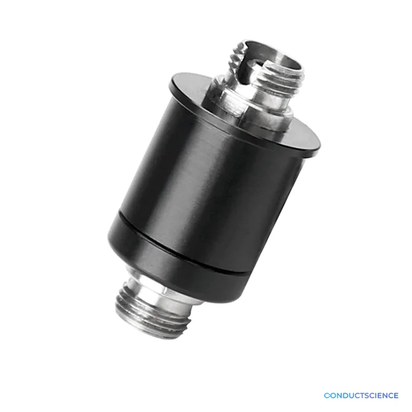
Dual Color Multichannel Fiber Photometry System
Fiber photometry recording systems record the activity of populations of neurons in specific brain regions through a calcium ion fluorescent indicator.
ConductScience offers Fiber Photometry System.

Louise Corscadden, PhD
Director of Science · ConductScience
Ask Louise about Dual Color Multichannel Fiber Photometry System fit, setup, configuration, or quote prep.
Already working with us? Sign in to connect this with My Scientist.
Key Specifications
Full details →- Model fit
- Mouse, Rat
- SKU family
- RWD-R810
- Sizing
- 34.0 x 39.0 x 33.0 cm
- Ordering
- Online checkout and quote request available
- Category
- Optogenetics & Photometry
- Build notes
- Confirm accessories, station layout, and support needs before purchase
Fiber optic recording systems record the activity of populations of neurons in specific brain regions through a calcium ion fluorescent indicator. In the study of neural circuits, the optical fiber recording system can stably monitor the group neurons of freely moving animals for a long time and then explore the correlation between neuronal activity and animal behavior.
The R810 dual-color multi-channel optical fiber recording system has two excitation light sources, 410nm, and 470nm, of which the unique 410nm can be used as the background signal to ensure the effective acquisition of real fluorescence data. 9 channels of data can be collected at the same time, which is suitable for simultaneous recording of multiple brain regions or multiple animals.
| Parameter Item | Specification |
|---|---|
| Exposure time setting range | 1-100 ms |
| Excitation wavelength | 410nm, 470nm |
| LED light power adjustment percentage | 0~100 % |
Introduction
Dual-color multichannel fiber photometry system is used for measuring in vivo neuronal activity in freely behaving animals. It can be used with genetically encoded calcium ion indicators (GECI) to measure the activity of groups of neuronal cell bodies, dendrites, and axonal terminals. Dual-colour photometry system comprises two excitation light sources that pass through a multimode optical fiber to stimulate neurons. The technique is frequently used to study neural circuit patterns and develop treatments for various neurological diseases, including traumatic brain injuries. Different neurons are simultaneously stimulated using different wavelengths of light.
Real-time monitoring of the neuronal activity at multiple sites in freely behaving animals is a bit challenging. It is impossible to record axonal terminals' activity using traditional electrophysiology because of the small sizes and high densities of these terminals, particularly in freely behaving animals. However, the situation becomes different when calcium imaging techniques are employed. For instance, two-photon (Ca+2) imaging allows monitoring in vivo Ca+2 activity at the micrometer scale and neuronal activity in superficial brain regions like the cerebral cortex. However, two-photon imaging is restricted to anesthetized or head-fixed animals. The development of miniaturized microscopes comprising gradient refractive index (GRIN) lenses allowed functional studies of deep brain regions (striatum, amygdala, and hypothalamus) in freely behaving animals. However, this technology also has a restricted recording range. Multichannel fiber photometry is far better than these techniques because one can easily monitor neuronal activity in many deep brain regions of freely behaving animals (Qin et al., 2019).
Principle
Fiber photometry is an optogenetics technique in which light is coupled into a multimode optical fiber and is directed toward the neurons. The light stimulates the neurons that emit fluorescent signals depicting the occurrence of neuronal activity. These fluorescent signals correspond to in vivo calcium dynamics, return through the same multimode fiber, and are captured by a detector. The fluorescent signal intensity changes from a "genetically encoded calcium indicator (GECI)" are detected. This way, calcium activity in different neuronal regions and neuronal circuit patterns are recorded. In many cases, multichannel fiber photometry systems are used. These systems allow recording neural activity in more than one brain region simultaneously. For instance, dual-channel fiber photometry systems have been developed that help record neural activity in two motor cortical regions or even two different animals simultaneously using a two-sensor photometry system.
Dual-color multichannel fiber photometry system works on the same principle described above, except that it employs two light sources for neuronal excitation. One excitation light source is used as a background signal to ensure the collection of authentic fluorescence data (Guo et al., 2015).
Apparatus and Equipment
Dual-color multichannel fiber photometry system offered by Conduct Science has two excitation light sources, i.e., 470nm and 410nm. The 410nm light source acts as a background signal for accurate real-time monitoring of the fluorescence data, whereas the 470nm light source excites the genetically encoded calcium indicators. The system utilizes nine channels to record neuronal activity simultaneously from multiple brain regions or multiple animals. The exposure time of the instrument ranges between 1-100ms, and its LED power can be adjusted between 0% and 100%.
Applications
Neuronal Population Dynamics Measurement
To understand the pathophysiology of Levodopa-induced dyskinesia (LID) in Parkinson’s disease (PD), Gao et al. (2022) studied the real-time activity of the Striatal D1 Dopamine neuronal population in rats. The researchers used a dual-color multichannel fiber photometry system to study the neuronal population dynamics. Heterozygous Drd1-Cre transgenic rats (age: 8-10 weeks, weight: 240 ±10gm) were selected as model animals. They ensured consistent genetic background, housed the animals on a 12-h light/dark cycle, and provided water and rodent chow ad libitum. After Levodopa administration following stereotaxic surgery of the subjects, the researchers used a dual-color multichannel fiber photometry system to detect and record fluorescent signals of a GECI named GCaMP6m. The scientists used a multimode optical fiber to guide light between the implanted fiber and the fiber photometry system. A light-emitting diode subjected lights of wavelengths 410nm and 470nm to excite Ca+2 -independent and Ca+2 -dependent control measurements, respectively. The emission light traveled through the same optical fiber was bandpass filtered and captured by a semiconductor camera. The scientists used a digital camera to record real-time rat behavioral videos. The data were statistically analyzed, and it was concluded that striatal D1+ neuronal population activity is linked to dyskinetic symptoms.
Measuring Long-Term Behavioral Changes resulting from Time-Restricted Feeding
Zhai et al. (2022) studied the effect of time-restricted feeding (TRF) on the long-term shift in locomotor behavior and the sleep-wake cycle in mice. The researchers used a dual-color multichannel fiber photometry system to compare intracellular Ca+2 signals in the GABAergic neurons of the suprachiasmatic nucleus (SCN) before and after TRF to analyze the activation of these neurons in dawn-TRF mice. The scientists took six to eight-week-old male C57BL/6J mice and housed them in cages with running wheels for 2 weeks under a light-dark cycle with food and water ad libitum.
After stereotaxic injections, a 200µm diameter optical fiber was implanted with 5 degrees in SCN in mice. The mice were screened for rhythmic Ca+2 signals using a multichannel fiber photometry system. The mice were kept in LD (light-dark cycle) for 3 days, subjected to TRF for 2 weeks, and then released into DD (dark-dark cycle) with ad libitum. Ca+2 signals were again recorded before TRF entrainment. The 470nm light source was used as a signal channel to stimulate neurons, and 430nm light was used as a negative control channel. The fluorescent signals were recorded every 35s at an optical power of 15-20 μw. The scientists concluded that in mice, SCN GABAergic neuron activity is linked to long-term behavioral changes (i.e., sleep-wake cycle and food-hunger responses).
Strengths and Limitations
Dual-color multichannel fiber photometry system has several advantages. The primary advantage of a multichannel photometry system is that it can record neuronal activity at multiple sites and in multiple animals simultaneously. In addition, the optical probe is small and flexible and causes minimum damage to the surrounding tissue (Wang et al., 2021). Furthermore, traditional electrophysiological recordings employ "optical tagging" to record the activity of cell-type-specific neurons. However, a major disadvantage of optical tagging is its sensitivity to noise interference in natural behaviors. On the other hand, the dual-color multichannel fiber photometry system is relatively efficient and not sensitive to noise interference because of utilizing genetically encoded Ca+2 indicators (Li et al., 2019). The technique is highly efficient and considerably less expensive. Other advantages of the system include high temporal resolution, high sensitivity, greater accuracy, and requires lower optical power.
Despite these advantages, a potential disadvantage of dual-colour multichannel fiber photometry system is that it has a poorer spatial resolution than other imaging techniques (Qin et al., 2019).
Summary
- Dual-color multichannel fiber photometry system is used for measuring in vivo neuronal activity in freely behaving animals.
- It can be used with calcium ion indicators to measure the activity of groups of neuronal cell bodies, dendrites, and axonal terminals.
- Dual-color multichannel fiber photometry system employs two light sources for neuronal excitation. One excitation light source is used as a background signal to ensure the collection of authentic fluorescence data.
- The system is employed to measure Ca+2 activity in multiple neuronal cell populations and multiple animals simultaneously.
References
Qin, H., Lu, J., Jin, W., Chen, X., & Fu, L. (2019). Multichannel fiber photometry for mapping axonal terminal activity in a restricted brain region in freely moving mice. Neurophotonics, 6(3), 035011.
Guo, Q., Zhou, J., Feng, Q., Lin, R., Gong, H., Luo, Q., ... & Fu, L. (2015). Multichannel fiber photometry for population neuronal activity recording. Biomedical optics express, 6(10), 3919-3931.
Gao, S., Gao, R., Yao, L., Feng, J., Liu, W., Zhou, Y., ... & Liu, J. (2022). Striatal D1 Dopamine Neuronal Population Dynamics in a Rat Model of Levodopa-Induced Dyskinesia. Frontiers in Aging Neuroscience, 14.
Zhai, Q., Zeng, Y., Gu, Y., Zhang, T., Yuan, B., Wang, T., ... & Xu, Y. (2021). Time-restricted feeding near dawn entrains long-term behavioral changes through the suprachiasmatic nucleus. bioRxiv.
Wang, Y., DeMarco, E. M., Witzel, L. S., & Keighron, J. D. (2021). A selected review of recent advances in the study of neuronal circuits using fiber photometry. Pharmacology Biochemistry and Behavior, 201, 173113.
Li, Y., Liu, Z., Guo, Q., & Luo, M. (2019). Long-term fiber photometry for neuroscience studies. Neuroscience Bulletin, 35(3), 425-433.
How It Works
Fiber photometry operates on the principle of bulk fluorescence detection from genetically encoded calcium indicators (GECIs) expressed in specific neuronal populations. Light-emitting diodes generate 410nm and 470nm excitation light that travels through multimode optical fibers to illuminate brain tissue containing calcium-sensitive fluorophores such as GCaMP. When intracellular calcium concentrations increase due to neuronal activity, the fluorophore undergoes conformational changes that alter its fluorescence emission properties.
The dual-wavelength design enables artifact correction by using 410nm as an isosbestic control wavelength where calcium binding does not affect fluorescence intensity. This reference signal accounts for movement artifacts, photobleaching, and changes in fiber coupling, while the 470nm channel reports calcium-dependent fluorescence changes. The system collects emitted fluorescence through the same optical fiber using a dichroic mirror and photodetector arrangement, with variable exposure times allowing optimization for different indicator kinetics and expression levels.
Nine independent channels enable simultaneous recordings from multiple fiber-optic cannulae, permitting multi-region analysis within single animals or parallel experiments across multiple subjects. LED power modulation (0-100%) allows precise control of excitation intensity to prevent phototoxicity while maintaining adequate signal-to-noise ratios for population-level calcium transient detection.
Features & Benefits
excitation_wavelengths
- 410nm, 470nm
number_of_channels
- 9
light_sources
- 2 excitation light sources
background_signal_wavelength
- 410nm
model_number
- R810
Automation Level
- semi-automated
Brand
- RWD
Research Domain
- Addiction Research
- Anxiety and Depression
- Behavioral Pharmacology
- Learning and Memory
- Motor Function
- Neuroscience
- Pain Research
- Social Behavior
Species
- Mouse
- Rat
Weight
- 8.27 kg
Dimensions
- L: 34.0 mm
- W: 39.0 mm
- H: 33.0 mm
| Feature | This Product | Typical Alternative | Advantage |
|---|---|---|---|
| Number of recording channels | 9 simultaneous channels | Entry-level systems often provide 1-4 channels | Enables multi-region circuit analysis and higher experimental throughput for complex behavioral paradigms |
| Excitation wavelength options | Dual wavelength (410nm, 470nm) | Single wavelength systems lack isosbestic control | Provides real-time artifact correction essential for freely moving animal experiments |
| Exposure time range | 1-100ms variable exposure | Fixed or limited exposure time ranges | Accommodates diverse calcium indicator kinetics and experimental temporal requirements |
| LED power control | 0-100% adjustable power | Fixed power output or limited adjustment range | Optimizes signal quality while minimizing phototoxicity for chronic recording applications |
| Isosbestic control wavelength | Dedicated 410nm isosbestic excitation | Varies by model - some lack artifact correction | Essential for robust calcium signal detection in freely behaving animals with movement artifacts |
The R810 combines 9-channel capacity with dual-wavelength excitation to provide both high experimental throughput and artifact-corrected signal quality. The inclusion of 410nm isosbestic control addresses a critical requirement for reliable calcium imaging in freely moving animals.
Practical Tips
Calibrate each channel using fluorescent microspheres or dye solutions before animal experiments to establish consistent baseline signals.
Why: Channel-to-channel variations can introduce systematic errors in multi-region comparisons.
Clean fiber optic connectors with appropriate solvents and inspect for damage after each experiment to maintain signal quality.
Why: Contaminated or damaged fiber surfaces significantly reduce light transmission and increase noise.
Record both wavelengths for at least 10 minutes before behavioral testing to establish stable baseline fluorescence levels.
Why: Initial fluorescence drift can confound calcium signal interpretation if not properly characterized.
Monitor the 410nm isosbestic signal throughout experiments to identify movement artifacts and verify fiber coupling stability.
Why: Large fluctuations in the isosbestic channel indicate optical artifacts that must be corrected during analysis.
If signals appear noisy, reduce LED power and increase exposure time rather than increasing excitation intensity.
Why: High excitation power causes photobleaching and phototoxicity that degrade signal quality over time.
Synchronize photometry recording with behavioral tracking systems using common trigger signals for precise temporal alignment.
Why: Accurate timing correlation between neural activity and behavior is essential for circuit function analysis.
Always verify LED output levels are appropriate for chronic recordings to prevent tissue damage from excessive light exposure.
Why: Phototoxicity can alter neuronal function and compromise experimental validity in long-term studies.
Calculate ΔF/F using the 410nm channel as the reference signal rather than using raw 470nm fluorescence values.
Why: Ratiometric analysis removes artifacts and provides more reliable calcium signal quantification.
Setup Guide
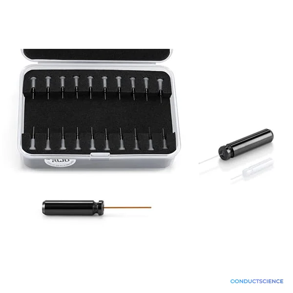
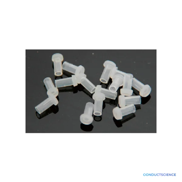
What’s in the Box
- R810 main control unit
- LED excitation modules (410nm and 470nm)
- Fiber optic input connectors (9 channels)
- Control software and drivers
- USB interface cable
- Power adapter and cables
- User manual and calibration procedures
- Fiber cleaning supplies (typical)
- Calibration standards (typical)
Warranty
ConductScience provides a comprehensive 1-year manufacturer warranty covering hardware components and LED modules, with technical support for system optimization and troubleshooting. Extended service plans are available for long-term research applications.
Compliance
References
Background reading relevant to this product:
What calcium indicators are compatible with the dual wavelength system?
The 410nm/470nm configuration is optimized for GCaMP variants (GCaMP6s, GCaMP7) and similar green fluorescent calcium indicators. The 410nm serves as isosbestic control where calcium binding doesn't affect fluorescence, while 470nm excites the calcium-bound state.
How does the 9-channel design improve experimental capabilities compared to single-channel systems?
Multi-channel recording enables simultaneous monitoring of interconnected brain circuits, bilateral region comparisons, or parallel experiments across multiple animals. This increases statistical power and allows investigation of coordinated network activity patterns.
What factors determine optimal exposure time settings for different experiments?
Exposure time (1-100ms range) depends on calcium indicator kinetics, required temporal resolution, and signal strength. Fast transients need shorter exposures (~10ms), while low-expression conditions may require longer integration times (~50-100ms) for adequate signal-to-noise.
How does LED power adjustment affect signal quality and tissue health?
Lower LED power reduces photobleaching and phototoxicity but may compromise signal-to-noise ratio. The 0-100% adjustment allows optimization for each preparation's depth and expression level while maintaining tissue viability for chronic recordings.
What is the maximum recording duration for chronic experiments?
Recording duration is primarily limited by calcium indicator expression stability and fiber implant integrity rather than instrument limitations. Typical chronic experiments span days to weeks depending on indicator choice and surgical quality.
How does artifact correction work with the dual wavelength approach?
The 410nm isosbestic wavelength provides a calcium-independent reference signal affected by movement artifacts, bleaching, and fiber coupling changes. Ratiometric analysis (470nm/410nm) removes these artifacts while preserving calcium-dependent signals.
What fiber optic specifications are required for optimal performance?
Standard multimode fibers (200-400μm core diameter, 0.39 NA) work effectively. Core diameter affects light collection efficiency and spatial resolution, while numerical aperture determines light-gathering capability from the tissue volume.
Can the system detect single-cell calcium events or only population activity?
Fiber photometry records bulk fluorescence from tissue volumes containing hundreds to thousands of neurons, providing population-level activity measures rather than single-cell resolution. For cellular detail, consult product datasheet for compatible microscopy options.
Have a question about this product?
Have a question? Just ask.
Send it over and we'll email you a personalized answer — no call, no scheduling.
Prefer to talk it through?
Accessories
Enhance your setup with compatible accessories